
NIBIB-funded researchers have developed a 3-D-printed scaffold coated in aggrecan, a native cartilage component, to improve the regeneration of cartilage tissue in joints. The scaffold was combined with a common microfracture procedure and tested in rabbits. University of Maryland researchers found the combination of the implant and microfracture procedure to be ten times more effective than microfracture alone. Microfracture alone is the standard therapy currently.
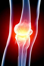
As people age, it is common for pain to develop in joints, especially in the knee joint. In 2017, reportedly close to twelve million individuals sought treatment for knee pain. Some knee pain problems arise from damaged articular cartilage, which is a rubber-like material that reduces friction in joints. Over time, articular cartilage can deteriorate or become damaged following injury or normal wear and tear, leading to the pain many people feel as they age.
Treating articular cartilage defects is challenging because cartilage tissue is non-regenerative, and implants poorly integrate with the native cartilage. One common procedure for cartilage restoration is the microfracture procedure, where damaged cartilage is removed, and small holes are created in the bone at the sites of cartilage removal. These small holes stimulate the growth of new cartilage by triggering the release of native mesenchymal stem cells (MSCs) from the bone. MSCs are the most vital factor for effective cartilage regeneration.
However, the microfracture procedure alone produces a weaker tissue in comparison to native cartilage because MSCs cannot easily locate or attach to the defect site(s). Other treatments can involve multiple surgeries or, depending on the level of damage, a total knee replacement. Lead researcher and director of the NIBIB-funded Center for Engineering Complex Tissues at the University of Maryland, John P. Fisher, Ph.D., sought to improve the quality of regenerated tissue during the microfracture procedure by developing a 3-D-printed scaffold. “Cartilage repair is a complicated research problem. Substantial progress will require a creative combination of methods and technologies to restore a material that was not meant to naturally regenerate,” said Seila Selimovic, Ph.D., director of the NIBIB program in Tissue Engineering.
“Our team, led by graduate student Ting Guo, Ph.D., who has since completed her degree, wanted to create a scaffold that could be readily translated into a clinical solution,” said Fisher. The scaffold, which can be printed in minutes, is functionalized with aggrecan to provide binding sites for cells released from the microfractures. The scaffold was tested in a well-established rabbit model for orthopedic surgery, where it was implanted over the defect site following microfracture. The scaffold guides MSCs and growth factors to the defect sites and strengthens cartilage regeneration.
Results from the study published in Biomaterials showed that the 3-D-printed scaffolds with aggrecan improved cartilage regeneration ten times more than microfracture alone or in combination with a non-functionalized scaffold. Congruently, aggrecan increased cell attachment to the scaffold by ten times in comparison to a non-functionalized scaffold—this was likely the reason why cartilage repair was improved. “Cartilage defects are a significant problem and can become a source of widespread arthritis and pain in our population. Our cartilage was not designed to function as long as we live now, so we need find ways to help it heal and improve quality of life,” says Jonathan D. Packer, M.D., collaborator and surgeon who performed the operations on the rabbits in this study.
In the future, Fisher and his team hope to optimize the functionality of the scaffold, so it selectively binds just MSCs rather than everything that gets released from the microfractures, to further strengthen the regenerated tissue. The next step involves replicating similar results to this initial study in a different animal model. Further down the road, Fisher would like to personalize each implant to a patient’s defect site by using readily available imaging already used in hospitals to pinpoint the exact size of the defect sites.
To read the research behind this article, please see: Ting Guo et al. 3D printed biofunctionalized scaffolds for microfracture repair of cartilage defects, Biomaterials (2018). DOI: 10.1016/j.biomaterials.2018.09.022
Read more at: https://medicalxpress.com/news/2019-01-bioactive-scaffolds-sore-knee-relief.html