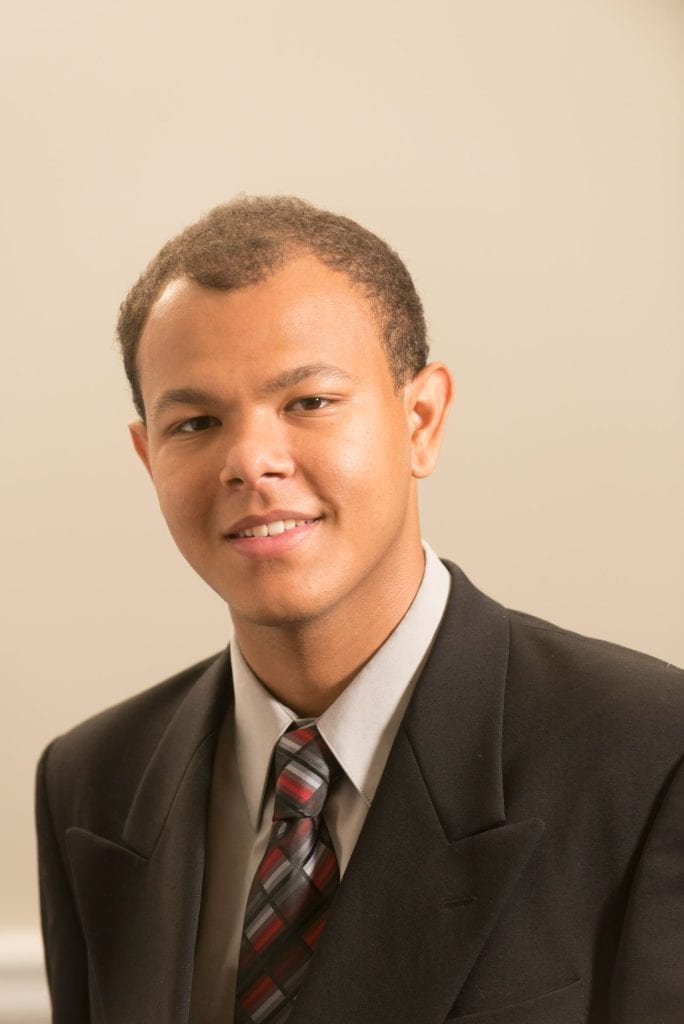
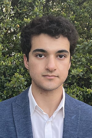
UMD Bioengineers Take New Approach to Engineering Heart Tissue


news story imageBIOE Professor Xiaoming (Shawn) He

University of Maryland professor Xiaoming (Shawn) He and fellow Fischell Department of Bioengineering (BIOE) researchers developed a new strategy to improve engineered heart tissues and one day advance overall understanding of heart disease.

According to the Centers for Disease Control and Prevention, heart disease remains the leading cause of death for both men and women in the United States. A major reason for this is that the human heart has very limited capacity to regenerate cardiomyocytes – the contractile cells of the cardiac muscle – when these cells become damaged. This damage commonly occurs due to ischemia – a reduction of blood flow to the heart, most often resulting from buildup or a blockage in the arteries.

Stem cell therapy is widely considered a promising strategy for treating heart disease. Given the significant ethical concerns associated with human embryonic stem cells, scientists look instead to use what are functionally embryonic stem cell-like cells – called human induced pluripotent stem cells (iPSCs) – to generate cardiomyocytes.

The science behind the process of tapping human iPSCs in this way is both fascinating and complex. For example, as an alternative to using human embryonic stem cells, bioengineers can take fibroblasts – the most common type of cell found in connective tissue – from a patient’s skin. They then “reprogram” these cells into iPSCs to convert – or differentiate – them into cardiomyocytes. To do this, historically, scientists have harnessed the power of viruses – such as adenoviruses – to transfer the genes they need into the harvested skin cells through the process of transduction. In this way, scientists can essentially “recode” skin cells to become cardiac muscle cells.

Unfortunately, the process poses many challenges. For one, the use of a virus or viral vector raises health concerns because of the potential for the virus to infect unintended cells in the human body. Even when scientists don’t use a virus in the reprogramming process, they face an additional hurdle. Believe it or not, iPSCs have the ability to transform into virtually any type of cell in the human body; this means that a human iPSC intended to be a cardiac muscle cell might become skin, hair, or bone instead.

Put simply: if the aim is to engineer cardiac muscle tissue, there is no room for error. A batch of human iPSCs intended for use in the heart simply cannot include an errant skin, hair, or bone cell.

But, the process of differentiating cells into cardiomyocytes can take time – as many as two weeks or more using traditional methods. In order to shorten the time frame and increase the yield of human cardiomyocytes, engineers often use 3D cell cultures rather than the conventional 2D approach. Even more, they use rho-kinase inhibitors – better known as ROCK inhibitors – to improve the survival of 3D-cultured human iPSCs before differentiating them into cardiomyocytes.

For years, engineers have accepted 10μM as the standard concentration of ROCK inhibitors for use in this process.

But years of hard work – and a little bit of luck – led Fischell Department of Bioengineering Professor Xiaoming (Shawn) He and a team of researchers to an important discovery. Their findings were published in Bioactive Materials.

He and his team found that the ROCK inhibitor commonly used in 3D cultures of human iPSCs contributed extensively to their heterogeneous differentiation. In other words, the ROCK inhibitor improved both the survival and yield of human iPSCs, but at a quality cost: the yields always included unintended cells, mixed in with cardiac muscle cells after cardiac differentiation.

“When put in 3D culture, human iPSCs easily undergo a type of cell death known as apoptosis, which can be minimized by adding 10μM ROCK inhibitor,” He said. “However, our work shows that this high concentration of ROCK inhibitor primes human iPSCs into not only the mesoderm for cardiac differentiation but also the other two germ layers –ectoderm and endoderm – for skin, hair, or bone differentiation, even before initiating cardiac differentiation.”

He and his team found that by reducing the concentration of the ROCK inhibitor to 1μM, they could dramatically improve the quality of human iPSC differentiation into cardiomyocytes. In fact, with this revised concentration, the team produced cardiac muscle cells with a beating efficiency of 95 percent in just a week, compared with when they used 10μM of the ROCK inhibitor; in the latter scenario, their yield of cardiomyocytes achieved a beating efficiency of under 50 percent in two weeks’ time. Even more, in the 1μM scenario, He’s team was able to synchronize the beating of their cardiomyocytes, creating a cardiac cell network – a cardiac spheroid – that beats at the same rate (~1 Hz) as that of the human heart. And, they did so with human iPSCs reprogrammed by a non-virus method.

“This paves the way toward producing a large number of highly pure and synchronized patient-specific cardiomyocytes for clinical use, with minimized concern on unintended tissue growth, arrhythmia, or viral infection,” He said.

Moving forward, He and his team hope to tackle the challenge of increasing their yield of 3D-cultured human iPSCs. Their work could help point to new techniques for large-scale production of human iPSCs-derived cardiomyocytes for use in engineering 3D cardiac constructs. Examples include bioprinting human hearts with therapeutic applications for heart attacks, heart transplantation, and high-fidelity drug screening of cardiotoxicity. The team’s work could also advance understanding of deadly heart diseases.

BIOE postdoctoral researcher Bin Jiang served as first author on the paper. In addition to Xiaoming (Shawn) He, BIOE postdoctoral researcher Wenquan Ou, Ph.D. students James Shamul and Samantha Stewart, visiting scholar Hao Chen, alumna Sarah Van Belleghem (Ph.D. ’21) and BIOE chair John P. Fisher also contributed to the paper, along with Zhengou Liu, M.D., Ph.D. of the University of Missouri’s Division of Cardiovascular Medicine.

Along with this work, He’s research group also develops novel strategies to bank human cells, tissues, and organs. Recently, they developed a novel natural sand-based technology to greatly improve the quality and yield of banking human iPSCs. This work is also published in Bioactive Materials.

In addition to his appointment as BIOE professor, He also holds affiliations with the UMD Marlene and Stewart Greenebaum Comprehensive Cancer Center and the UMD Robert E. Fischell Institute for Biomedical Devices.

Published February 11, 2022


Bioactive scaffolds guide sore knee relief, cartilage repair

NIBIB-funded researchers have developed a 3-D-printed scaffold coated in aggrecan, a native cartilage component, to improve the regeneration of cartilage tissue in joints. The scaffold was combined with a common microfracture procedure and tested in rabbits. University of Maryland researchers found the combination of the implant and microfracture procedure to be ten times more effective than microfracture alone. Microfracture alone is the standard therapy currently.

As people age, it is common for pain to develop in joints, especially in the knee joint. In 2017, reportedly close to twelve million individuals sought treatment for knee pain. Some knee pain problems arise from damaged , which is a rubber-like material that reduces friction in joints. Over time, articular cartilage can deteriorate or become damaged following injury or normal wear and tear, leading to the pain many people feel as they age.

Treating articular cartilage defects is challenging because cartilage tissue is non-regenerative, and implants poorly integrate with the native cartilage. One common procedure for cartilage restoration is the microfracture procedure, where damaged cartilage is removed, and small holes are created in the bone at the sites of cartilage removal. These small holes stimulate the growth of new cartilage by triggering the release of native  (MSCs) from the bone. MSCs are the most vital factor for effective cartilage regeneration.

However, the microfracture procedure alone produces a weaker tissue in comparison to native cartilage because MSCs cannot easily locate or attach to the defect site(s). Other treatments can involve multiple surgeries or, depending on the level of damage, a total knee replacement. Lead researcher and director of the NIBIB-funded Center for Engineering Complex Tissues at the University of Maryland, John P. Fisher, Ph.D., sought to improve the quality of regenerated tissue during the microfracture procedure by developing a 3-D-printed scaffold. “Cartilage repair is a complicated research problem. Substantial progress will require a creative combination of methods and technologies to restore a material that was not meant to naturally regenerate,” said Seila Selimovic, Ph.D., director of the NIBIB program in Tissue Engineering.

“Our team, led by graduate student Ting Guo, Ph.D., who has since completed her degree, wanted to create a scaffold that could be readily translated into a clinical solution,” said Fisher. The scaffold, which can be printed in minutes, is functionalized with aggrecan to provide binding sites for cells released from the microfractures. The scaffold was tested in a well-established rabbit model for orthopedic surgery, where it was implanted over the defect site following microfracture. The scaffold guides MSCs and growth factors to the defect sites and strengthens cartilage regeneration.

Results from the study published in Biomaterials showed that the 3-D-printed scaffolds with aggrecan improved cartilage regeneration ten times more than microfracture alone or in combination with a non-functionalized scaffold. Congruently, aggrecan increased cell attachment to the scaffold by ten times in comparison to a non-functionalized scaffold—this was likely the reason why cartilage repair was improved. “Cartilage defects are a significant problem and can become a source of widespread arthritis and pain in our population. Our  was not designed to function as long as we live now, so we need find ways to help it heal and improve quality of life,” says Jonathan D. Packer, M.D., collaborator and surgeon who performed the operations on the rabbits in this study.

In the future, Fisher and his team hope to optimize the functionality of the , so it selectively binds just MSCs rather than everything that gets released from the microfractures, to further strengthen the regenerated tissue. The next step involves replicating similar results to this initial study in a different animal model. Further down the road, Fisher would like to personalize each implant to a patient’s defect site by using readily available imaging already used in hospitals to pinpoint the exact size of the defect sites.


To read the research behind this article, please see: Ting Guo et al. 3D printed biofunctionalized scaffolds for microfracture repair of cartilage defects, Biomaterials (2018). DOI: 10.1016/j.biomaterials.2018.09.022

Read more at: https://medicalxpress.com/news/2019-01-bioactive-scaffolds-sore-knee-relief.html

TEBL Study Led by Dr. Ting Guo Published in Biomaterials

A new study written by a team from the Center for Engineering Complex Tissues (CECT) will soon be published in the journal Biomaterials. The study looks into the process of developing a 3D-printed aggrecan functionalized scaffold in order to aid with microfracture procedures. The study’s aggrecan functionalized scaffold showed better improvement of regenerated cartilage tissue than those treated with traditional methods or left untreated. The study was a collaboration between Dr. Jonathan D. Packer from the Department of Orthopaedics at the University of Maryland School of Medicine in Baltimore and departments within the University of Maryland in College Park, MD. Other researchers included Ting Guo, Maeesha Noshin, Hannah Baker, Evin Taskoy, Sean J. Meredith, Qinggong Tang, Julia P. Ringel, Max Lerman, Yu Chen, and John P. Fisher.

While millions of Americans are impacted by cartilage defects, the current treatment (Microfracture and Autologous Chondrocyte Implantation) has many post-surgical difficulties and a long recovery time. This method involves drilling the layer of bone just beneath the cartilage defect to release mesenchymal stem cells (MSCs). This often leads to a weaker regenerated tissue than healthy cartilage. The new aggrecan functionalized scaffold method used in the study resulted in histologically healthier and thicker cartilage tissue formation compared to the original method.

After further examination, the team found that the aggrecan functionalized scaffold led to higher cell adhesion than control groups. For the first time in the field, the team also evaluated the cartilage regeneration at a functional level through a newly designed locomotion test. Despite this, the aggrecan functionalized scaffolds had a more even distribution of newly formed cartilage that was significantly thicker than the other groups. It also showed better chondrocytes growth and ECM formation.

When asked about the impact of the study, lead author Ting Guo said, “The presented biofunctionalized acellular scaffold combined with microfracture shows promise for clinical translation. Such acellular technologies stand to greatly impact future clinical solutions to improve the quality of repaired cartilage tissue as demonstrated in this study without additional surgeries and with a relatively low cost compared to cell-based therapies.”

This study showed that using an aggrecan functionalized scaffold can lead to better support for people undergoing treatment for articular cartilage defects, as demonstrated by improved tissue quality. This provides another possible and more efficient treatment method to study and utilize for the millions of patients affected by articular cartilage issues.  

For more information, the original article can be read on Science Direct. The study has also been highlighted by Science Translational Medicine, which can be accessed here.

Charlotte Piard Speaks at Conference in Belgium

On Friday, October 12, 2018, TEBL member Charlotte Piard gave a presentation at the 3rd annual 3D Printing and Bioprinting in Healthcare Conference in Brussels, Belgium. The presentation was entitled “Biomimetic Cell-Laden 3D Printed Scaffolds for Bone Tissue Engineering.” During the presentation, Charlotte presented her research as well as an overview of other TEBL lab research topics. The conference gathers 3D printing industry leaders, business heads, medical consultants, researchers and engineers from across the globe. More information about the event can be found here.

Javier Navarro Speaks at IX Seminary on Biomedical Engineering in Colombia

Javier Navarro giving his presentation at SIB in Colombia

TEBL member Javier Navarro spoke at the IX Seminary on Biomedical Engineering (SIB) at the Universidad de Los Andes in Bogotá, Colombia. The event took place May 16 – 19, 2018. Javier’s talk was entitled Assessment of the Effects of Energy Density in Crosslinking of Keratin-Based Photo-Sensitive Resin.”

The presentation was about keratin and the creation of a resin designed for 3D printing keratin. This resin was then used to determine how much energy projected during curing defines different properties of keratin hydrogels. Navarro and other TEBL members Jay Swayambunathan, Marco Santoro, and John P. Fisher found that energy density regulated the chemical reaction needed to crosslink keratin and determines how much the hydrogels crosslink. This, when taken together with its saturation nature, determine microstructural properties of the hydrogel. By understanding the role of energy density in creating the crosslinked network, they were able to adjust the printing parameters of the resin in a cDLP 3D printing system.

For more information, the IEEE proceedings article can be found here.